

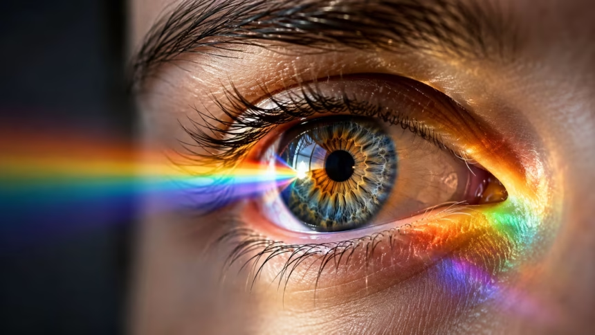
How Does the Human Eye See Colors?
Have you ever wondered why a strawberry looks red, the sky appears blue, or a leaf seems green? Color feels like a natural property of the world around us, but it is actually an experience created by the eyes and brain. Objects do not contain color in the way they contain shape or weight. Instead, they reflect different wavelengths of light that the visual system interprets as particular colors.
Understanding how the human eye sees colors requires looking at several connected processes. Light must first reach an object, reflect from its surface, enter the eye, and strike specialized cells inside the retina. These cells convert light into electrical signals, which then travel through the optic nerve to areas of the brain responsible for visual processing and color perception.
The human visual system can distinguish millions of different colors by comparing signals from several types of light-sensitive cells. However, people do not always experience color in exactly the same way. Genetics, age, lighting conditions, eye health, surrounding colors, and brain processing can all affect how a shade appears to an individual viewer.
Learning how color vision works can help explain everyday experiences, including why colors look different under artificial lighting, why some people are color-blind, and why optical illusions can confuse the brain. It also reveals that seeing color is not a simple camera-like process. It is an active interpretation created through cooperation between light, the retina, nerves, and the brain.
What Is Color and Where Does It Come From?
Color begins with light, which is a form of electromagnetic energy. The electromagnetic spectrum includes radio waves, microwaves, infrared radiation, ultraviolet radiation, X-rays, and gamma rays. Human eyes can detect only a small portion of this spectrum, known as the visible light spectrum. This visible range contains the wavelengths that people commonly experience as red, orange, yellow, green, blue, indigo, and violet.
Each visible color is associated with a general range of wavelengths. Longer visible wavelengths are usually perceived as red or orange, while shorter wavelengths appear blue or violet. Green and yellow fall between these ranges. However, wavelength alone does not completely determine color perception because the brain compares multiple visual signals rather than simply labeling each wavelength separately.
When light strikes an object, some wavelengths may be absorbed while others are reflected. A green leaf appears green because pigments in the leaf absorb many wavelengths while reflecting more light from the green portion of the visible spectrum. The reflected light enters the eye, allowing the visual system to create the experience of green.
The amount and type of light available also influence the color that reaches the eye. A shirt may look bright blue in daylight but slightly dull or purple under indoor lighting. This happens because different light sources produce different combinations of wavelengths. Color is therefore created through an interaction between the light source, the object’s surface, the eye, and the brain.
How Light Enters the Human Eye
The process of seeing color begins when reflected light enters the eye through the cornea. The cornea is the clear, curved surface covering the front of the eye. It provides much of the eye’s focusing power by bending incoming light toward the interior structures. Because the cornea must remain transparent, injury, swelling, or disease in this area can interfere with clear vision.
After passing through the cornea, light travels through the pupil, the dark opening in the center of the iris. The iris is the colored portion of the eye and controls the size of the pupil. In bright conditions, the pupil becomes smaller to reduce the amount of incoming light. In darker conditions, it becomes larger so that more light can enter.
Light then passes through the lens, which fine-tunes the focus before the image reaches the retina. Small muscles adjust the shape of the lens depending on whether a person is viewing something near or far away. This focusing process helps produce a clear image, although the image projected onto the retina is upside down and reversed.
The retina is a thin layer of light-sensitive tissue lining the back of the eye. It contains millions of photoreceptor cells that respond to light. Once these cells are stimulated, they begin converting light energy into electrical and chemical signals. These signals form the foundation of both brightness detection and human color vision.
The Role of Rods and Cones in Color Vision
The retina contains two primary types of photoreceptor cells: rods and cones. Rod cells are highly sensitive to low levels of light and support vision at night or in dim environments. They are excellent at detecting movement and changes in brightness, but they do not provide detailed color information. This is why the world appears less colorful in very dark conditions.
Cone cells are responsible for most color vision and fine visual detail. They work best in bright or moderately lit conditions and are concentrated in the central area of the retina. Cones allow people to notice small details, read text, recognize faces, and distinguish between closely related shades. Without functioning cone cells, normal color perception would not be possible.
The highest concentration of cone cells is found in the fovea, a tiny area near the center of the retina. When a person looks directly at an object, the eyes position its image on the fovea. This creates the sharpest and most detailed part of the visual field. Peripheral vision contains fewer cones and more rods, making it better for noticing movement than identifying precise colors.
Rods and cones work together to create a useful visual experience across changing conditions. In daylight, cones dominate and produce detailed, colorful vision. As lighting becomes dimmer, rods become more active while cone sensitivity decreases. The gradual shift from cone-dominated vision to rod-dominated vision explains why colors fade as evening turns into darkness.
How the Three Types of Cone Cells Detect Color
Most people have three main types of cone cells, which is why human color vision is described as trichromatic vision. These cone types respond most strongly to different ranges of visible light. They are commonly called short-wavelength, medium-wavelength, and long-wavelength cones, or S-cones, M-cones, and L-cones.
S-cones respond most strongly to shorter wavelengths associated mainly with blue and violet light. M-cones are most sensitive to medium wavelengths associated broadly with green. L-cones respond strongly to longer wavelengths connected with yellow, orange, and red. However, each cone type reacts to a wide and overlapping range rather than detecting only one specific color.
The brain determines color by comparing the relative activity of these three cone types. For example, yellow light may strongly activate both L-cones and M-cones while producing relatively little activity in S-cones. Blue light creates stronger activity in S-cones, while certain green shades produce greater stimulation in M-cones. Different combinations create the enormous variety of colors people can recognize.
This comparison system explains how the eye can perceive colors that are not represented by a single wavelength. Purple, for instance, is created when the visual system receives a combination of red-related and blue-related signals. There is no single purple wavelength in the visible spectrum. The perception is produced by the pattern of cone stimulation and the brain’s interpretation of that pattern.
How the Brain Turns Light Signals Into Color
Photoreceptors do not send finished color images directly to the brain. Instead, cone cells pass information to several layers of retinal neurons, including bipolar cells and ganglion cells. These cells organize, compare, and modify the signals before they leave the eye. Important processing therefore begins inside the retina rather than waiting until the information reaches the brain.
The axons of retinal ganglion cells join together to form the optic nerve. Each optic nerve carries visual information from one eye toward the brain. Some nerve fibres cross at a structure called the optic chiasm, allowing information from both eyes to be combined. This arrangement helps the brain build a unified visual field and support depth perception.
Visual signals then travel through several brain regions before reaching the visual cortex at the back of the brain. The visual cortex analyzes features such as edges, shapes, movement, brightness, orientation, and color. Different groups of neurons respond to different visual patterns, allowing the brain to construct a detailed interpretation of the scene.
Color perception is therefore not produced by the eyes alone. The eyes collect light and convert it into neural signals, but the brain gives those signals meaning. Memory, attention, expectations, context, and previous experiences may influence the final result. This is why two people can look at the same image yet describe its colors somewhat differently.
Why the Brain Uses Opposing Color Channels
Color processing involves more than comparing three types of cones. The nervous system also organizes color information into opposing channels. According to the opponent-process system, some neurons compare red-related signals against green-related signals, while others compare blue-related signals against yellow-related signals. A separate channel processes differences between light and dark.
These opposing channels help the brain detect color contrast efficiently. A neuron associated with the red-green channel may become more active when red-related signals are stronger and less active when green-related signals dominate. This comparison allows the visual system to identify subtle differences between nearby objects and separate them from their backgrounds.
Opponent processing also explains why people normally do not see a color that is simultaneously reddish-green or bluish-yellow. Red and green are processed as opposing directions within one channel, while blue and yellow form another opposing pair. Although colors can be mixed physically, the brain’s neural coding places certain experiences on opposite ends of the same system.
The same process contributes to negative afterimages. After staring at a bright red object for an extended period, the red-sensitive part of the visual system temporarily adapts. Looking at a white surface afterward may create a greenish afterimage because the opposing green-related response becomes relatively stronger. Similar effects occur with blue and yellow combinations.
Why Colors Change Under Different Lighting
A color can appear different depending on the strength, direction, and quality of the surrounding light. Sunlight contains a broad range of visible wavelengths, while artificial lights may produce an uneven spectrum. Traditional warm bulbs often emphasize yellow and orange wavelengths, whereas cooler lights may contain more blue-related wavelengths.
The surface of an object also affects how it reflects light. Smooth, glossy materials may create bright highlights, while rough surfaces scatter light in several directions. Transparent, metallic, fluorescent, and matte materials interact with light differently. As a result, two objects made with the same pigment may not appear identical when their textures or finishes differ.
Despite changing conditions, the brain often maintains a relatively stable impression of an object’s color. This ability is called color constancy. A white sheet of paper usually continues to look white outdoors, indoors, or in a shadow, even though the exact wavelengths entering the eye have changed. The brain estimates the lighting conditions and adjusts its interpretation.
Color constancy is helpful but not always perfect. Unusual lighting, photographs, digital screens, and strong shadows can mislead the visual system. Viral images in which viewers disagree about the color of a dress or shoe demonstrate this effect. Different brains may make different assumptions about the light source, producing different color experiences from the same image.
How Surrounding Colors Affect What We See
The appearance of a color is influenced by the colors placed around it. A medium-grey square may seem lighter against a black background and darker against a white background. The square itself has not changed, but the brain compares it with the surrounding brightness. This process is known as simultaneous contrast.
Nearby colors can also shift the perceived hue of an object. A neutral shade may look slightly green when surrounded by red or slightly yellow when placed beside blue. Designers, photographers, artists, and marketers use these relationships to make particular colors appear brighter, warmer, cooler, softer, or more noticeable.
The visual system relies on contrast because absolute measurements of light would be less useful in everyday life. Lighting changes constantly as people move between rooms, shadows, outdoor spaces, and digital displays. By comparing one area with another, the brain can recognize objects and boundaries more reliably despite these changes.
However, contrast-based processing can also create optical illusions. Patterns, shadows, gradients, and background colors may cause identical areas to look different. These illusions do not mean the eyes are failing. They reveal the shortcuts and comparison methods the brain normally uses to create fast, stable, and meaningful visual perception.
Why Some People Experience Color Blindness
Color blindness, more accurately called color vision deficiency, occurs when one or more cone systems do not function in the usual way. The condition does not always mean that a person sees only black and white. Most affected individuals can still see many colors but may have difficulty distinguishing specific shades or color combinations.
Red-green color vision deficiency is the most common form. It usually involves differences in the L-cones or M-cones and can make reds, greens, browns, and oranges harder to separate. Because genes associated with common red-green deficiencies are located on the X chromosome, the condition is more frequently inherited by males.
Blue-yellow color vision deficiency is less common and affects the ability to distinguish certain blues, greens, yellows, and reds. Complete color blindness is rare and may involve serious problems with cone function. People with severe forms may experience poor visual sharpness, light sensitivity, and limited or absent color perception.
Color vision changes may also develop later in life due to eye disease, nerve damage, certain medications, chemical exposure, or neurological conditions. A new or sudden change in color perception should not automatically be considered inherited color blindness. It may require assessment by a qualified eye-care professional to identify the underlying cause.
How Age and Eye Health Affect Color Perception
Color perception may gradually change as people grow older. The lens inside the eye can become thicker and more yellow over time, filtering some shorter wavelengths before they reach the retina. This can make blue and violet shades harder to distinguish and may cause certain colors to appear less vivid.
The number and efficiency of retinal cells may also change with age. Older adults may need brighter lighting to identify subtle color differences, particularly when contrast is low. These changes usually happen slowly, meaning a person may adapt without immediately noticing that their color discrimination has become less accurate.
Eye conditions can affect color vision more significantly. Cataracts may alter brightness and color balance by clouding or yellowing the lens. Disorders involving the retina or optic nerve can reduce color sensitivity, create washed-out vision, or make one eye perceive colors differently from the other. The specific effect depends on which visual structures are involved.
Regular eye examinations can help identify changes before they seriously affect daily activities. People should seek professional advice if colors suddenly appear faded, distorted, unusually dark, or different between the two eyes. Prompt assessment is especially important when color changes occur alongside blurred vision, eye pain, flashes, blind spots, or other new symptoms.
Can Humans See Every Possible Color?
Human eyes can distinguish a remarkable number of colors, but they cannot detect every form of electromagnetic radiation. Infrared light has wavelengths longer than visible red, while ultraviolet light has wavelengths shorter than visible violet. These forms of radiation exist around us, but the normal human visual system does not interpret them as visible colors.
Other animals may experience color differently because their photoreceptor systems are not identical to ours. Some birds, insects, fish, and reptiles possess additional cone types that respond to ultraviolet wavelengths. Certain mammals have fewer cone types and may see a more limited range of colors compared with most humans.
Digital screens also cannot reproduce every color the human eye might encounter. Screens create colors by combining red, green, and blue light at different intensities. Printers usually rely on cyan, magenta, yellow, and black inks. Each device has a limited color gamut, meaning some real-world shades cannot be reproduced perfectly.
There may also be small differences in the range of colors different people can distinguish. Most humans have three functional cone types, but variations in cone pigments and neural processing may affect sensitivity. A small number of people may possess additional cone variations that could support unusually fine color discrimination, although the practical experience can vary considerably.
How Does the Human Eye See Colors in Everyday Life?
In everyday life, color vision helps people recognize objects, read signals, select food, notice hazards, understand emotional expressions, and navigate complex surroundings. The brain performs these tasks so quickly that most people rarely notice the many stages involved. Light collection, cone activation, retinal comparison, nerve transmission, and brain interpretation occur within fractions of a second.
Color also interacts with attention and memory. Bright or contrasting colors may attract the eyes more quickly, while familiar color patterns can help people recognize brands, traffic signs, maps, and household objects. However, emotional reactions to colors are influenced by personal experience and culture rather than being determined only by the biology of the eye.
Understanding color perception is especially useful when choosing lighting, designing digital content, creating accessible websites, or presenting important information. Relying on color alone can create difficulties for people with color vision deficiencies. Clear labels, strong contrast, patterns, icons, and readable text make visual information easier for everyone to understand.
Ultimately, answering “How does the human eye see colors?” involves much more than identifying red, green, and blue cone cells. Color is a carefully constructed experience produced through the interaction of visible light, photoreceptors, retinal circuits, the optic nerve, and multiple areas of the brain. What people see is not simply light itself, but the brain’s useful interpretation of it.
The Science Behind Color Vision Explained
The human eye sees colors by detecting patterns of visible light reflected from objects. Cone cells in the retina respond to overlapping ranges of wavelengths, while rods support vision in dim conditions. The visual system then compares these responses to identify differences in hue, brightness, and saturation.
Signals from the retina travel through the optic nerve and reach specialized processing areas inside the brain. These regions evaluate color alongside shape, contrast, movement, lighting, and surrounding objects. The brain also uses opposing red-green and blue-yellow channels to organize color information more efficiently.
Because perception depends on both light and interpretation, colors may appear different under changing conditions. Shadows, artificial lighting, surrounding shades, age, genetics, and eye health can all influence the final experience. Color constancy usually helps the brain keep familiar objects looking stable, but unusual images can occasionally challenge that system.
Human color vision is therefore an active biological process rather than a direct recording of the outside world. The eyes gather visual information, but the brain constructs the experience people recognize as color. This remarkable partnership allows humans to distinguish subtle shades, understand visual signals, appreciate art, and interact safely with their surroundings.
Frequently Asked Questions
What part of the eye allows us to see color?
Cone cells in the retina are mainly responsible for color vision. They detect different ranges of visible light and send signals that the brain compares to create the experience of color.
Why can we not see colors clearly in the dark?
Cone cells need relatively strong light to function effectively. In darkness, rod cells become more active, helping with movement and brightness detection but providing little meaningful color information.
How many colors can the human eye see?
The human visual system may distinguish millions of color variations under suitable conditions. The exact number depends on lighting, contrast, eye health, genetics, and how differences between colors are measured.
Do both eyes see colors in exactly the same way?
The two eyes usually perceive colors similarly, but minor differences can occur because of lighting, lens clarity, retinal sensitivity, or eye health. A sudden or major difference between the eyes should be professionally assessed.
Is color created by the eyes or the brain?
Both structures are essential, but the final experience is created by the brain. The eyes detect light and convert it into signals, while the brain compares and interprets those signals as recognizable colors.